Brief #
50 nanograms of almost anything injected is plenty for mass spectrometer systems.
we get a protonated molecule, M+H or a deprotonated molecule. They are not molecular ions that’s what you get in an EI spectrum. They’re not protonated molecule ions that’s redundant. A protonated molecule is an ion basically.
Jack doesn’t like to call it a chromatogram.
The alternative technique of atmospheric pressure Chemical Ionization is also a useful technique for certain molecules. This one is most useful for very polar molecules, and macromolecules, peptides and proteins and unlike those have nitrogens. They are can be protonated. They’re easily to form them in an aqueous solution. This technique is not amenable. APCI is not good for proteins or inorganic or large molecules. It is much better for nonpolar molecules, steroids, for example, and other kinds of nonpolar molecules.[23:45]
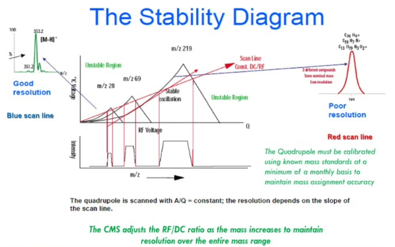
We do not see ion current unless it’s above this Scan Line. And as you move it up, you can see that you’re going to have less than less of this peak, and that’s increasing the resolution. The Scan Line determines the resolution.
Good practice: I trust it, but I take a look at it first thing in the morning, I run something I know the result. You want accurate mass with measurement to the nearest 10th. so checking on our daily or certainly weekly basis. And certainly minimally every month, you should do a mass axis calibration and an auto tune to make sure that it’s it’s correctly running.
At higher and higher mass, the baseline resolution becomes more challenging.
When ions hit the quadrupole, they are lost, therefore the sensitivity is poor.
I would go back at the beginning and clean to clean the source, or re tune it to make sure we’re getting good ion current performance, rather than kill my multiplier.
calibrate and tune mass spectrometer can optimize peak shape.
There’s another way of defining molecular weight that I’d like you to forget about because this is not the answer that the mass spectrometer is it’s called the relative atomic mass.
The low mass region below mass to charge 150 has a lot of chemical noise.
If you show me a mass spectrum, I can tell instantly whether you know what you’re doing and are doing a good job of mass spectrometry or haven’t tuned your system in a long time, I look for isotopes in the correct ratio.
Ions go in this serpentine path in quadrupole analyzer.
APCI technique: liquid was introduced through the capillary to the chamber. Liquid turns to plume. A discharge needle is placed in the plume. We put 4000 volts on it. Gas phase molecules are ionized by gas phase chemistry.
What does autotune do?
) mass accuracy, resolution (FWHM - full width half max), peak symmetry, intensity.
) Tuning raises/adjusts the scan line of the quadrupole mass analyzer. Its goal is to get/maintain resolution according to the stability diagram.
Disadvantage of LC/MS comparing to GC/MS: The mobile phase, water, acetonitrile, methanol, maybe some additives like formic acid has a lot of chemical noise in the low mass region below mass to charge 150. Methanol adducts with itself.
Advantage of LC/MS comparing to GC/MS: you can see real molecule.
Transcripts #
… let me get started on the first lecture. So understanding atmospheric pressure ionization API can stand for the American Petroleum Institute, or active pharmaceutical ingredient. In this class, it stands for atmospheric pressure ionization. And 30 or 40 years ago, that was unheard of term, we never made ions at atmospheric pressure, we made ions in the vacuum system, if you have any experience with electron ionization, or chemical ionization and GCMS ions are made in inside the vacuum not in the in the atmosphere. So that’s what’s unique about what we’re doing. So some of this is pretty basic for someone like yourself, but let’s start this all out in the same plane by understanding what a mass spectrum is.
A mass spectrometer has several components, we’ll talk about each of them in some detail. We have to make ions in the first place, gaseous ions, and that therefore, we have a source for those ions and ions sources. We have those ions go into the vacuum system after they’re formed at atmospheric pressure into the mass analyzer. The ions are detected the at the end the exit of the mass analyzed by a detector, electron multiplier.
And of course, a vacuum is important. Why is the vacuum important in mass spectrometry? if you remember your organic chemistry, ions need to have a mean free path 1, a long mean free path. If we have ions that atmospheric or they run into gas molecules, they run into nitrogen and oxygen, whatever other gases in the system. In a slide going through some of these carnival experiences for kids go through and they hit bags and so forth, it’s not a clean path for the (xxxxx, not clear) is a vacuum. So we need to mean free path of several meters, by having a vacuum 10 to the minus six tore (atmospheric pressure being 760 Tore), then we can have a mean free path of several meters on our ions don’t hit anything. They can go where we want them to go. So a UV detector does not have to have a vacuum, a gas chromatograph FID detector, you don’t have to have a vacuum. But mass spectrometry must have a vacuum for the ions to get where we want them to go.
And so if we have that, we have to vacuum pumps, and we’ll talk about each of that. The mass spectrum is shown in the bottom here, it’s an XY plot, x is the mass to charge ratio, the mass x and the ordinate or y axis is the percent abundance. So a mass spectrum has a base peak, there should be only one base peak. If you have more than one base peak, you have saturated the detector, and everything’s off scale. And by the way, mass spectrometers, including ours are very sensitive. And I am on my favorites comments is the solution to pollution is dilution. We don’t inject micrograms of a sample, these things are really sensitive, 50 nanograms of almost anything injected is plenty for ours and other mass spectrometer systems to use.
Organic chemists, synthetic chemists make milligrams, sometimes grams of material. And they really need to dilute that reaction mixture dramatically. They must be you must be teaching them from time to time to dilute the samples before they inject.
And so we have a mass spectrum with peaks mass-to-charge ratios, if you will. The most abundant one is called the base peak. And the others are relative to it the relative relative abundance on the ordinate. And so here’s a deprotonated line in this example a fragment ion and another fragment ions, and that’s a fingerprint for that organic molecule. And so that’s the main thing that we often are looking for. With that fingerprint, we can begin to identify or characterize the molecule that we’ve just analyzed.
Molecular Weight information is certainly available from a mass spectrometer. And the question here is why do we combine HPLC with mass spectrometry? The most common thing someone wants a synthetic chemist is the molecular weight. And it’s important to note with these atmospheric pressure ionization techniques, we do not get the molecular weight itself directly. we get a protonated molecule, M+H or a deprotonated molecule. I’m a little bit of a picky guy nomenclature and so they are not molecular ions, that’s what you get in an EI spectrum. They’re not protonated molecule ions that’s redundant. A protonated molecule chemists knows is an ion basically. So we are getting molecular weight indication via protonated molecule or deprotonated molecule. And as you probably know, and we’ll see later, we can have other kinds of adducts, a proton adduct is an adduct, we can have a methanol adduct, we can have an M+32, if we will or M+33. And so solvent ion molecule reactions can occur. Most of the time, it’s fairly clean in electrospray, that we have a protonated molecule with another ionization technique that we’ll talk about, which has atmospheric pressure chemical ionization, we have fewer adducts. We’ll talk about that in a later lecture in this session.
I like to suggest that the mass spectrometer is the truth machine, students used to call it a truth machine. That’s why they’re so important and forensic applications if you watch certain TV programs. And I was just set a meeting on forensic toxicology, or police work, other kinds of work on drug identification, it’s very important to have proof, good selective information that the molecule reporting is not just the retention time. If you do a GC-FID, you have a retention time and you can inject co-elute something and have it be the same retention time, but you can still be fooled. Another molecule may have that same retention time under those conditions, it’s very unlikely that another molecule will have the same retention time and the same mass spectrum, because chromatography separates out isobaric molecules, those with the same molecular weight. So it’s the ultimate selectivity.
It’s really useful for confirmation of HPLC peak purity, you probably know that UV detectors can now can be diode array. It’s a scanning UV spectrum and some people feel that Scot significant or acceptable selectivity. It’s better selectivity to just 254 nanometers, but it still is not as good as a mass spectrometer. If you can have a diode array detector in line with a mass spectrometer, nothing wrong with that. That’s added selectivity. It’s very useful for characterization of isomeric mixtures including chiral mixtures. Mass spectrometry is a chiral. It cannot distinct or differentiate chiral molecules but coupling chromatography with it, HPLC, even GC, you can actually separate enantiomers which are mirror images of each other. And so it’s very good for that.
And it’s very important for use of quantitation. Now the original business that I co-founded was a CRO doing LC-MS/MS for the pharmaceutical industry. It’s right next door. It’s called it used to be called ad Mian but it’s been acquired by a company called Quintiles, this time called Q squared solution, there are 65 mass spectrometers over there serving the pharmaceutical industry. And we use stabilized label isotope molecules to do quantitation. Originally, old timers think of mass spectrometry just for qualitative analysis. In the 60s and early 70s. It was used just for identifying things. It can still be used for that. But an awful lot of mass spectrometry has been sold not for qualitative analysis, but quantitative analysis. How much drug in terms of nanograms per ml is in this clinical sample? And so quantitative analysis is really a big business in LC-MS and LC-Ms/Ms. And the part The unique aspect of an isotopically labeled internal standard is it is chemically identical to the D0 or the unlabeled molecule. And it absolutely col-elutes with a molecule but its mass is different. If it’s a D3, got three deuterated atoms in it, its masses, three mass units higher in the mass spectrometer can easily distinguished that’s a powerful combination, and is revolutionized pharmaceutical analysis. So LCMS with for quantitation is very important. And can be done with our Advion compact mass spectrometer.
Now if we get into some of the details of our instrument, our instrument looks like this. It’s the vertical as opposed to horizontal. Sometimes we wish we’d made it flat. So you could stack it like some others do, but it’s not. One of the important thing that we are proud of with our system is that we have a very good vacuum system, we have a rough pump and a turbomolecular pump. And as my opening comment said, if you want to do good mass spectrometry, you need a good vacuum. There are some systems on the market without mentioning any names that have tried to be to make it very inexpensive and very small and very light. And they have diaphragm pumps, simple pumps, the kind of thing you’ll have in an aquarium, if you will. And those cannot create the vacuum that makes for a good mass spectrometer.
And so here’s a cross section of our system. There’s the box horizontally inside. we hope to give you a tour later our manufacturing is right nearby. And at some point during your visit here it will show you the insides of these things where they’re made right around the corner. And so we have a sprayer we used to say we would spray and pray that would work that was in the 70s and 80s. And we did spray and pray that it would work. Now we don’t have to pray at least on this anymore, we spray and we know how to spray and make it make a good series of ions.
So we have a sprayer in all of LCMS you’ve got a liquid and we need to make tiny droplets. And we’ll talk more about that. So this is an atmospheric pressure, we have ions that are sampled into this capillary into this region. And we have a pumping system for that region, they go through the ions makeup turn, it’s really helpful to have ions have a nonlinear path. Ions are I like to say somewhat like teenagers, you can sort of guide them, you keep them out of trouble, if you will. And so ions we can make to make the turn but neutral molecules of which there are a lot that are not affected by the electric fields and they can be pumped away. So it’s nice to have ions make a turn of some sort. And most commercial systems on the market today have some form of an ion path that’s nonlinear basically. And so we spray we have a sub ambient region here and as we go further along this path, we have higher and higher vacuum, if you will. So the sample introduction can be done by HPLC, by infusion, by flow injection analysis,
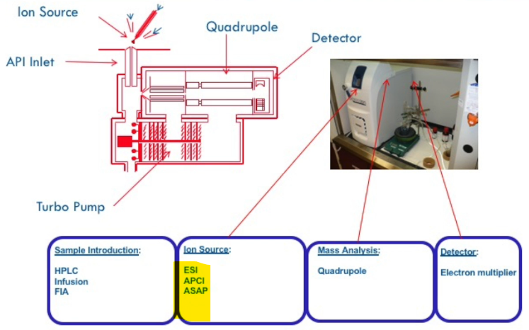 we’ll talk about each of these, the ion source make science either by electrospray atmospheric pressure, chemical ionization, or another approach that we call ASAP that we’ll talk about a simple technique that it does not use HPLC as an inlet system. By the way, there are almost there’s approaching 30 different ways of making ions that what the vast majority are by electrospray or APCI. The others are more esoteric and developing. The mass analysis designed by a quadrupole. We’ll talk more about that. That’s this section right in here. And the detector is an electron multiplier at the end of the ion path. And we’ll talk about each of these components in a little detail in sequence. I like to be very analytical. My wife says I’m anal. I say no, I’m analytical and take this in a very systematic way.
So this is a slow video of of our system. If I click on an issue start to see the spray come down through the capillary, the voltages are shown here, the droplets are evaporated, and electrostatically focused in the capillary. And they make the turn here, neutrals being pumped away by the vacuum pump. Here are the pressures or vacuum in these areas regions, they continue through this, this ion optics to the quadrupole mass analyzer. And we’ll talk about these more in a bit. But the only selected or filtered ions are passed through the detector, and they go to the detector and that gives us our mass spectrum. It gives us an ion chromatograph profile. If you’re doing LC that’s called a chromatogram. And ion chromatogram. Some people call things chromatograms. When there was no LC, again, I’m picky. I don’t like to call it a chromatogram. If there was no chromatography, there is another term some people use a [xxx], I’m not sure where that came from. But in any case, we get ion current profile. sample coming off and going away in the mass spectrum or the fingerprint.
So the next slide shows the details of that, if you will. With respect to the various voltages, when you go in the laboratory, I hope that you can think like a mass, I think like a lot like an ion. And by the way, I have an insight edge on this, if you take note of my last name, a student well into my career pointed out to me that I have ion in my last name, no wonder I like this field, I guess. So a Henion doesn’t really exist. But it launches, I want you to think like an ion and, what would it be like to go through here and be in see the detector, again, we’ll talk about each of these components in sequence.
So the vacuum system very important to have a good backup system, our system can be put on a cart, there is a stackable part, whether it’s an entire LCMS system on the cart, and on the bottom is this vacuum pump. And people don’t like this vacuum pump. Because it’s heavy, it makes a little bit of noise, it’s a really good vacuum pump, it’s a rough pump. vacuum is created by two stages of pumping. The first stage is a rough pump or rotary pump. And we’ll talk about this one. And that pumps the vacuum down to 10 to minus 2, 10 to minus three tour. But we need a better vacuum than that. That’s where the turbo molecular pump comes in, that we will talk about in more detail in a bit. The two pumps together make a very good vacuum system. And our system really is well pump. So the rubbery pump or rub pump is that one on the bottom is connected by a hose to the inlet or to a whole pipe on the back of the or mass spectrometer. And the turbo molecular pumps are shown here. This is a unique one with differential pumping across it. And these are old rings that bolt up underneath, you never have to worry about this part, these are very reliable. They’re also very expensive, you’re going to have to buy one of these. But this is the heart of the system that creates the high vacuum, there’s creates a ultra clean vacuum system and the regulation and maintenance of the vacuum is all done by the by the system.
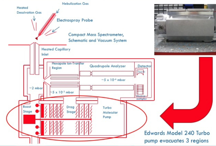 So here is again, a cross section of our mass spectrometer the mass analyzer region, this is what the component the whole component looks like when it’s out of the system. What I want to point out here is that we have three regions of evacuation, there is the first region there’s the second region and there’s the third region. this one turbo pump pumps all three of those regions. This is called the boost stage, the drag stage and the turbo like molecular pump, you don’t need to know or worry about that. But it’s useful to know that one pump is doing a really nice job of pumping this whole analyzer region. And that’s a little unusual, other commercial systems in the past have had two or three different turbo molecular pumps, each one dedicated this region one dedicated to this region and so forth that adds the cost that adds the complexity and we cannot take too much credit other than being smart enough to choose this because this is a commercially available pump that can be that others use and we have chosen to use for this system.
 So we
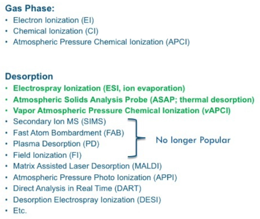
So we spray into this capillary make ions and that atmospheric pressure in the pumping system maintains 10 to minus six tors, or there abouts. Ionization we need to make ions, all mass spectrometry makes a gas phase ion. If we make the ion at atmospheric pressure, we’ve got to somehow get it into the vacuum system and into the mass analyzer. Historically, electron ionization used to be called something called electron impact, but it was never really an impact. But the correct term is electron ionization that’s still in vogue. It’s still very popular for GCMs gas chromatography mass spectrometry. You can also do ChemiCal ionization inside of a for GCMs. And we’re not doing either of those. There’s also atmospheric pressure chemical ionization and we and others can do that for LCMS. There are other the all those ions are under the gas phase incorporated into a gas phase.
Other techniques in involve invoke what’s called desorption ionization. And there are a variety of them. Some of them are shown down here. But electrospray is essentially a desorbing. The ion is formed in a droplet, and we’re going to see the droplet gets smaller and smaller and smaller. But at some point, the ion is desorbed from that droplet, it’s called I like this term, it’s called ion evaporation, it literally evaporates from the droplet into the mass spectrometer, and that’s the heart of electrospray as we’ll see. The other techniques that I’ve shown in screen that we use on our system available to you is this atmospheric solids analysis probe. This is a simple way of introducing a sample it does not involve LCMS in any way. It says probe that you put your sample on, and you’ll see this operate in the laboratory. And of course, we have vapor atmospheric pressure chemical ionization, or vAPCI, see, as we call it, which you’ll learn about too. other techniques are commercially available. I might add that these FAB Fast Atom Bombardment used to be a detergent, I don’t know if it still exists. But these are no longer commercially available. These were brought out in the late 70s 80s and have come and gone basically, they’ve been put out of business by newer, better ionization techniques. MALDI matrix assisted laser absorption is alive as well, that’s still a very popular technique, we do not provide MALDI for our system. And these other techniques, Atmospheric Pressure Photo Ionization, DART, DESI, and this etc, includes another 25 Different ionization means of doing that. But the most popular across the board are electrospray and APCI techniques.
Slide: The Ion Source #
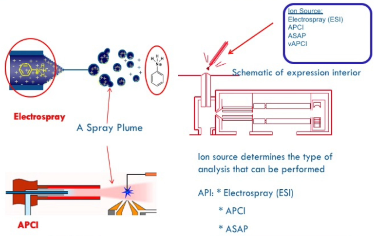
[21:41] So the ion source, how do we make ions? We have two major ways I’ve just mentioned electrospray, I have also mentioned APCI. So let’s look at electrospray. This is a very narrow or capillary, think of a syringe needle, maybe a 10 microliters syringe needle, and let’s convert that into what we call a sprayer, we’re going to have liquid going through it, pumped by either an infusion pump or an LC pump. So the inside diameter is very small, probably 100 microns, what’s the diameter of a human hair 80 microns. So this capillary, this needle is very small inside diameter, liquids going through it, and we're going to put high voltage on it. This goes back to 1984, when John Fenn, who shared the Nobel Prize several years ago, reported this. develop this. It actually goes back to 1967, a researcher at in Chicago, who did the very first electrospray, but did not appreciate how powerful it could be. It was John Fenn, who applied it to macromolecules and small molecules. And that was the beginning of a revolution. I was of interest in the mid 80s. That was 1984. In the mid 80s, I followed his work at my research group, and we were quickly sold on this, I like the idea, you couldn’t buy it, you had to make it yourself, no big deal. And we did. And I reported it at a ASMS meeting in 1985. I went to that was in June, and in October, I went to another meeting in Germany and an agent at the time of that ASMS meeting, there were four people on the planet, we were one of them doing electrospray. By November, there were 20 people doing it, it just took off nothing took off any faster than the interest in electrospray because John Fenn showed that it could do proteins and in even inorganic polymers.
So electrospray makes these droplets, ions are in the droplets, in solution if you will. And it undergoes the ion evaporation model 2, which we’ll talk about more right in the region of this inlet to the mass spectrometer. ions are formed that go into there in our mass analyzer. The alternative technique of atmospheric pressure Chemical Ionization is also a useful technique for certain molecules. This one is most useful for very polar molecules, and macromolecules, peptides and proteins and unlike those have nitrogens. They are can be protonated. They’re easily to form them in an aqueous solution. This technique is not amenable. APCI is not good for proteins or inorganic or large molecules. It is much better for nonpolar molecules, steroids, for example, and other kinds of nonpolar molecules [not clean about this part].
And unfortunately, in my view, this is not used anywhere near as much as it could. I don’t know how often you use it in your training with your students. I would suggest it should not be forgotten. The vast majority people use only electrospray evens under some conditions or doesn’t work that well and APCI would work better. Think of it as a tool in your toolbox. A mechanic or a plumber takes out the optimum tool for whatever they’re doing. There may be times when you’re struggling having electrospray work when APCI might work better. The unique difference between electrospray and APCI is we have this discharge needle, call it a spark plug, it’s a needle with high voltage three or 4000 volts. It creates literally a spark and that’s positioned in the plume, the vapor that’s coming through this system. there is no such needle - discharge needle in electrospray. The ions are formed by applying voltage to this capillary itself, they aren’t the voltage is applied to this needle in this case, and these plume has gas phase molecules in it. And they are ionized by gas phase chemistry. So, this is in solution chemistry, this is gas phase chemistry a big difference between the two. If gas phase chemistry is required for APCI, you can sort of understand why myoglobin [a protein] will not work well by APCI. It’s 9,000, like your wages doesn’t have a vapor pressure, you’re not going to be able to do a protein with APCI. So, these are both useful tools, you may have purchased only one or the other, or you may have purchased both, it’s up to your applications. So, that’s an intro to these two major ion sources that are available.
Types of mass spectrometers #
Types of mass spectrometers, ours is a single quadrupole mass spectrometer. there are triple quadruples, and by the way, the more the Q’s you have, the more the price goes up. So instead of 50, 60, $70,000, they can be $500,000. If you want to get an orbittrap down here, it can take me three quarters of a million dollars. These get much more expensive, easily over $500,000. But these are powerful analytical tools. And in the CRO next door, I think we had four orbitraps and some ToF. Another instrument that’s used to be more popular than it is now at the moment, there’s only one vendor that sells FT-ICR Fourier Transform mass spectrometry. That’s Bruker. It used to be the thermo had one, but they came thermo came up with the orbitTrap. It is so powerful that they stopped analytically powerful that they no longer sell a FTMS. So these are other kinds of mass analyzes. The system you have from us is a single quadrupole.
The Quadrupole Mass Analyzer #
So the quadrupole mass analyzer, if you break the vacuum, shut the pumps off and vent the system, this sometimes needs to be done, you look inside, you can see the quadrupole assembly as shown here. So this familiar schematic shown here, the ion source, the ion optics in the beginning and the quadrupole, we are looking at this region right here. And at the end exit of the quadrupole is the electron multiplier detector.
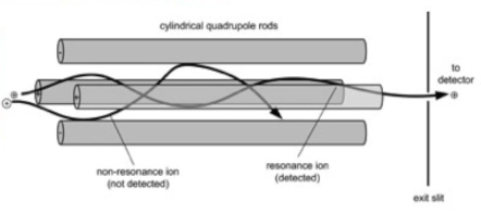 Ions go through the quadrupole. I initially naively thought ions went through like a laser, just like a light beam, they don’t. they go with us in the three dimensional Serpentine, if you will. path is shown here. There’s a software called SIMION. It reproduces the paths of ions with computers. And it shows that these ions go in
 this serpentine path. Our goal, your goal is to have make sure the ions don’t hit the surface of the rod, because then they’re lost. So mass resolution ion optics is keeping the ions going through the center of the quadrupole and resolving them. And we’ll talk more about that later.
So the ions go through under different conditions under different conditions with different RF, radio frequency DC voltages, and adjacent ions are resolved. So they all get to the detector at different times. And that composite view is a mass spectrum of all those ions.
The Stability Diagram #
Note: It is about the Scan Line.
[28:19] So this is a little bit technical, but I always found this very helpful. You have auto tune, and you can go in and manually tune the system. And I would encourage you to learn a little bit more beyond auto tune so you can appreciate what’s going on. In the properly adjusted mass axis, you we see ions that are above this red line, this is called a Scan Line. It’s a constant DC RF voltage applied by the system. And we do not see ion current unless it’s above this red line, this red line can be displaced upwards at the same slope. And as you move it up, you can see that you’re going to have less than less of this peak. And that’s increasing the resolution, we increase the resolution as we raise this Scan Line. And we can raise it or lower it with a constant slope. Or we can alternately hold this origin the same and just rotate this up or down. And you can see that that would affect the high mass end, this is the high mass end if you will, the more than the low mass end and so autotune does all that. And if you really get good at this and and want to optimize your system, some customers like to go in and manually do it and you can do that. So the Scan Line determines the resolution.
This is an example of good resolution 353.23 And that’s 350.3. So they are they are baseline resolved or maybe 5 or 10% generally resolved. That’s at the low end, it’s easier for a quadrupole to have unit mass resolution. That’s what set is called as unit mass resolution at the low end. Our quadrupole goes to 2000 mass units, and others do too. And any quadrupole has more of a challenge they can do it, but maintaining good resolution high mass at the upper end is more challenging. And so here is an example of poor resolution, not from our system, just as for didactic purposes are three ions in there, and they’re not resolved, you would have to increase the slope of this Scan Line. And if you did, you could then resolve those ions. So that’s what you’re doing when you’re increasing the resolution, the mass resolution, I’m not talking about chromatographic resolution, is sort of analogous, but it’s mass resolution, and auto tune and you are in or your own hands. If you have peaks that are not hopefully this bad off, but maybe you see a shoulder they’re barely resolved, you’re going to improve that by increasing the slope of this line. And that’s called the stability diagram.
The CMS (Compact Mass Spectrometer) adjusts the RF/DC ratio as the mass increases to maintain unit mass resolution across the mass range. The quadrupole must be calibrated using known mass standards. We infuse or inject a known mixture of compounds where we know the masses and we calibrate the mass axis and the resolution based upon that compound. And then when you run unknowns, you can be confident you will get the correct mass. Do you have a Mettler balance in your laboratory, Fisher balance and Accurate balance? If you’re, if you’re well run managed laboratory you probably have that someone come in every six months or so to calibrate that. you don’t just use a mass Mettler balance for 10 years straight without over a mass of adjusting it. And so a mass spectrometer is similar in that needs to be calibrated. Mass access calibration adjusted on an ongoing basis. You don’t have to do it every day. I’m really picky. And I really want to be confident I trust but verify. It wasn’t Reagan that made that popular. I trust it. But I take a look at it first thing in the morning, I run something I know the result. And I make sure those masses are correct. And that’s not shift, the mass axis isn’t shifting. If you have a really bad day, or you never haven’t adjusted our resolution in months, it’s possible for it to drift instead of being 302.3. It could be 302.6, or eight or even worst case, a mass unit off, you want accurate mass with measurement to the nearest 10th. And so checking on our daily or certainly weekly basis. And certainly minimally every month, you should do a mass axis calibration and an auto tune to make sure that it’s it’s correctly running. Don’t just trust it like a UV detector, the only thing you have to worry about UV detector is the light going out. And you will know because you’ll see nothing. On this one, it takes a little more attention to make sure that it’s behaving itself.
Slide #
[32:48] So here’s what we typically show for Windows, these are zooming in on certain regions. This is the wiring diagram or schematic for, you don’t have to worry about this taking care of running in the quadruples, by the way the quadruples are about that long, I think you’ll see some they’re about as big as that a little thicker. They’re highly precisely made. We don’t make them, very few people make them. It takes real skill to do that. They are must be absolutely accurately arrayed. It’s a quadrupolar array for them. And they are not just stuck together with glue. They’re very, very precisely made. And so here’s a mass range, this is 74. Going out the 52. It’s hard to see this 1034 Think out here. And you can see this is 74. This is 75 well resolved masses. This is 200 and something in the one the next mass you know, so you want to see pictorially like this that the adjacent masses differing by one are easily resolved. They don’t have to be widely resolved. You don’t have to drive cars between them. But you want to have them baseline resolved as shown here. And as you can see at higher and higher mass, the baseline resolution becomes more challenging, but this is perfectly adequate for typical for most applications.
Slide: Cartoon #
Here’s a cartoon or a video showing how ions go through the mass analyzer. Notice they don’t go through like a straight line. If they are lost, if they aren’t focused correctly, they’ll be lost and that means you have poor sensitivity, you means you haven’t not resolved the mass resolution well enough to have it. Get the ions into the detector.
Slide: Mass Spectrometer Components - Detector #
The other mass mass mass spectrometer components the one most importantly are very important at the end exit of the mass exit at the exit of the quadrupole is a electron multiplier detector. I like that term because it is a multiplier. It has a gain of million. 10 to the six. that means that one ion hits the surface, here comes an ion into the surface, And that produces an electron, remember it’s an electron multiplier. This does not measure directly ions, it creates a beam of electrons. So one ion hits the surface, and makes the electron come off. I will have a cartoon later, a movie showing that. that hits the surface and two electrons come off, those two ions sit here and it cascades. And that happens a million times very, very quickly. So the ion current through the quadrupole is infinitesimally small weak, it must be amplified by a million to actually see our signal. So there’s not lots of ions going through at one time on the quadripolar, there are a few and we amplify that in any mass spectrometer, operates in the same way. So very weak ion current exiting the quadrupole. But that gets amplified through this approach by electron multiplier.
Slide: Cartoon of Electron Multiplier Operation #
So here’s a cartoon of that. this is kind of slow, but it shows an ion coming in to hit this surface. It happens much faster than that. now there’s an electron coming off, they hit that surface, that comes down here, creates two electrons. And that continues on and on, amplifying it, happening very very fast. We can [xxx] 1000 1000 1000s of Dalton’s per second, and this can keep up with it. And so that’s the electron multiplier at the end of the exit. This does not last forever. This is a consumable, you need to appreciate that once in a while, maybe once a year, if you have take good care of it, and don’t run it at too high of voltage. In my last two years or more, some people start to see deterioration of performance, poor sensitivity. And so they jack up the voltage on the multiplier, that hurts it, that’ll give you more sensitivity, but I would go back at the beginning and clean to clean the source, or retune it to make sure we’re getting good ion current performance, rather than kill my multiplier. So just a point in noting that the multiplier is something that from time to time, you must purchase a new one and have it installed. And either do that yourself or hire that done by someone who is used to doing that.
Slide: calibrating and tuning the mass spectrometer #
calibrating and tuning the mass spectrometer, something we don’t have to do with an FID detector or UV, it’s very important to calibrate in tune that’s tuning the ion and optics adjusting it for optimum sensitivity to ensure that the mass accuracy the resolution, and the peak symmetry and intensity are all within instrumental specifications, it can get off its feet, it can start to look poor as a result of not doing a recent tune. it can go weeks and weeks without doing it but you cannot go forever without doing it. A mass calibration is performed to adjust the measured mass to charge ratio to match the reference mass-to-charge with a with a allowable tolerance of $\pm$ 0.1. So we do not have accurate mass measurement accurate mass measurement, you need an orbit trap or a time of flight to have 0.1562 really an accurate mass, but we can readily provide $\pm$ 0.1 mass units we and other quadruples. autotune is performed with just the measured mass resolution and full width of half max is this term to be within allowable limits and the universe resolution at half width halfway up the peak 0.5 - 0.7 m/z (m over z) unit says generally defined as unit mass resolution those adjacent masses will be near baseline resolved as well to optimize the peak shape. They the ions when I say peak here, it’s ion peaks not chromatographic peace and intensity of the caliber and peaks Caliburn peaks are the reference compound that we’re using. There’s two vials one for positive ion, one for negative ion and you infuse those in, it’s a mixture of chemicals and we you know, the masses in the system can be calibrated based upon those, it’s critical that the system is calibrated in tune to encompass the analytes mass to charge ratio that is an analyte m over z must lie within a calibration mass range for mass spectral accuracy.
Slide: Resolution Calculation #
So here is full width of half mass these are two adjacent masses, these are not chromatograms. This is a mass to charge ratio. And at full full width at half max. If that is 0.5 - 0.7 Dalton’s as defined as unit mass resolution. That’s the kind of performance you should get. resolution as long been defined as m over delta m - the mass the actual mass divided by that delta mass. And that’s the resolution. How we define and others define mass resolution in a mass spectrometer.
Slide: Interpretation of Mass spectra Caffeine #
So let’s take good old caffeine, there is this elemental composition C8H10. High resolution is needed to accurately get that in elemental composition. There are some software and other ways of doing that. But in general, call it single quadrupole does not give us that but it does give us the molecular weight. So the nominal mass so this molecule is eight times 12, eight, eight times carbon, 10 times hydrogen. It’s nominal mass, not as accurate mass, but its nominal basis four this for nitrogen, so four times 14 and so forth. That adds up to 194 molecular weight. There’s another way of defining molecular weight that I’d like you to forget about because this is not the answer that the mass spectrometer is it’s called the relative atomic mass. And I was horrified when I first learned that the Merck index, you can look at the Merck index, which has a list of lots of drugs by molecular weight, and they have list them listed in there as relative atomic masses. This is before the Merck goes way back to the 1800s. This is befor mass spectrometry. And I’ve seen people going into pharmaceuticals few years ago, and they would have a Merck compound and they would, they would adjust their mass spectrometer to get the answer that Merck index had rather than what how God made the molecule. And so the what you really want is what’s shown here, the Monoisotopic exact mass. So this is the accurate mass hydrogen is 1.0078. Roughly point one, what what this is, is the isotope weighted mass, so a weights the percentage of carbon 13, the weight of them weights, the mass of carbon 15, these are small amounts that shifts the mass higher, generally those masses are higher in mass, that’s 0.09, the real answer is 0.08, are you really bothered by that, perhaps not. But a higher molecular weight molecule, when you get the four, and five, and 800, that becomes much more different. And so the monoisotopic get exact mass is what you should be adding up. And that’s what your mass spectrometer when properly tuned and calibrated will give you.
Slide #
[41:28] So here’s a mono.., this is mass-to-charge. Looks like a chromatogram. But as always pay attention to the x axis, here is your molecule, there is the 195.1. If we go back one, if you round this up to the nearest 10th, so 194.1, and you add a proton, and you have 195.1, it’s not off by one, that’s because we’ve added a proton. That’s how we form an ion for caffeine. Importantly, what really brought electrospray on board was its ability to do large molecules. And so here is a one mass unit apart from these ions, this is a fairly high molecular weight here is 1340, 1349, or 48, that’s 48, 49, 50. These, the distance between these, which is something you should pay attention to is one mass unit. What’s the difference between these two, this is a doubly charged peptide, the difference here is a half a mass unit. So m over z is simple arithmetic, the mass of protonate, a molecule divided by one gives you this spacing here, if you divide by two, you get half of that. And so the spacing between ions is a signpost for whether it’s a doubly or triply charged ion. It’s hard to distinguish beyond that by looking at the mass spectrum, there’s software that can do that. But be aware that if you have a doubly charged ion, that means a peptide has two protons on it rather than just one. Some peptide is going to have three protons. a protein 150 kilodaltons can have hundreds of protein of protons on it. And so they’re multiple charged ions with these are called. any of you looking at peptides and proteins, it’s certainly capable of doing that. small molecules, you don’t have to worry about this.
Slide #
A representative API mass mass spectra, assured molecular weight, as long as you can do earth or arithmetic, the molecular weight is not this peak, it’s one less than that, because that’s M+H. So I facetiously say you need to know how to subtract or add one one. If you have some fragmentation occurring, then you can have product ions or fragment ions. And we’ll talk more about that. This is not MSMS we’ll show you how that if you have some in source fragmentation called spread to the fragment or bolded. Some companies call it that we can take this mild ionization, electrospray and APCI is very mild ionization, the ionization itself does not cause fragmentation. It’s very gentle, unlike electron ionization, which is 70 electron volts and lots of energy. But we can increase the energy of an ion and cause or produce fragmentation to get more information.
Slide #
Positive ion LCMS, some examples, here’s malathion. That’s the chromatogram coming out in a little over five minutes. If we put the cursor on that peak, we can get its mass spectrum. Here’s the mass spectrum always pay attention to the axis as as m over z. This is time. And so we can look at the mass spectrum of any peak, there’s only one here. This is the protonated molecule. Here’s a fragment ion. And if you’re one thing I didn’t ask is how many of you have a basic understanding of organic chemistry, Garden Wright organic chemistry, we’re talking about molecules that are organic molecules, you all have a comfortable feeling good. That’s really helpful to understand what’s going on here because we like to speculate or postulate what this ion might be in this isn’t a toxic group. And if you protonate that you can lose ethanol. And that’s that’s a good a plausible explanation for why we’re seeing this product. I’m here that makes perfect sensor that could happen that would be a stable oxonian[??] ion. So that’s that example.
Slide: Comparison between Full-Scan API and API SIM #
comparison between full scan, atmospheric pressure ionization and selected ion monitoring, a lot of people call this single ion. If you’re only monitoring one, I can agree that sing single line, but the correct term is selected ion monitoring. And so here’s our information on our software, we have clicked, we’ve got a 20 minute run we’ve we’ve told it to do to scan the scan range, which is from 100 to 700. And we’re doing that this scan rate. And when we do that, we get this mass spectrum, we get these ions that are that are labeled, if we instead want to do selected iron monitoring, which is targeted analysis, that was you’ll see later that that can be used for quantitation, Or for improved sensitivity, we can ask the mass spectrometer or tell it using SIM, we click this button instead of this one, you’ll see this in the lab. And we can select 609, 472 and 311 that we’re going to monitor and we see this abbreviated mass spectrum, that’s SIM acquisition. So those are two very important acquisition profiles that you can use.
Slide: Selected Ion Monitoring (SIM) #
Selected ion monitoring monitors only selected ions from the full scan mass spectrum. So you have to know what the mass spectrum is first, right? You cannot do this on a total unknown molecule because you don’t know what the ions are going to be. You can speculate but you may be wrong. This technique does not scan the mass spectrometer but rapidly jumps from selected ion to selected ion jumps through that space. It’s used again added sensitivity and selectivity over a full scan mass spectrum. It has been said that you’re going to be it can be up to 100 times more sensitive than full scan. So if you’re having trouble seeing something on their full scan opposition get a little bit LC peak. If you’d switch to SU and selected ion monitoring reinject select the ions of interest, you’ll see great big nice pretty LC peak, when you couldn’t see very well before. It’s a dramatic improvement in sensitivity and selectivity.
It is used for targeted analysis, select an ion a minor thing means you have to know what you’re looking for and what those ions are.
In the favorite applications where this is used, certainly for quantitative analysis with a single mass analyzer system. And by the way, if you have a stable isotope internal standard, but D3, or six C13 is an aromatic ring. This is a golden way of doing quantitative analysis that the system can be done a selected ion monitoring, and you’ll alternately monitor the D0 versus a D3 or there’s the C12 versus a C13 isotopes.
increase sensitivity and reduce interference from chemical noise, the signal to noise ratio will be different. One elephant in the room dirty little secret we have, we’re doing LCMS. And what’s in the mobile phase, water, acetonitrile, methanol, maybe some additives like formic acid, the low mass region below mass to charge 150 has a lot of chemical noise, ethanol methanol adducting with itself. The good news is if we want to look at a real molecule a molecule we might better you do GCMS, we LCMS for intractable molecules that you don’t want to make derivatives of that, that are higher mass. And so in generally, when you scan full scan, you do not scan down to mass-to-charge 20, huge chemical noise, we usually scan, hope assuming our molecule, we’re not going to see something below 150, I like to use 150 as the low end, I think the example I just showed you was mass-to-charge 200, 200 to 700, or something like that. The reason we don’t scan full scan is because there’s chemical noise down there in an in trying to see the command LC chromatogram for a molecule that will be underneath the chemical noise. Take that one step further and do selected ion monitoring. Now you’ve got like to say blinders on the mass spectrometer, you’re not looking at chemical noise at all, it’s there, but you’re only looking at the signal of interest, and your signal to noise will be dramatically improved. Which sensitivity one definition is signal to noise.
Slide #
Here’s some practical examples of Enlite herbicides is a DuPont herbicide, they’re using soybeans. And here’s a mixture of three of them. And you click on or touch the cursor on that peak. And you get that mass spectrum with the isotopes nicely shown. And if you show me a mass spectrum, like I’m showing here, I can tell instantly whether you know what you’re doing and are doing a good job of mass spectrometry or haven’t tuned your system in a long time, I look for isotopes in the correct ratio, I don’t want this eight plus one to be half of that or higher than that that’s that’s representative of how many carbon 13 are in there, which is only 1.1% for each carbon. And so for each of these LC peaks we get a fingerprint of these molecules. This one happens to have looks like it has some chlorine in it doesn’t quick glance I can see a three to one ratio with the chlorine isotope.
If that was full scan, if I now go positive ions selected ion monitoring, I see the the total selected ion that’s what this is a total Select ion current, here is this molecule. And we have two of them with the same same mass and we’ve got one that’s called eluding and we got one that’s separated here. So selected ion monitoring on this would be for I’m going to show you a soil extract. If you want to look for pesticides, herbicides in soil, then you would you select an ion monitoring, and that looks like this. So here’s a farmer spraying his soybeans in the field before he plants it. And here are these drugs are compounds herbicides from the soil. And you can readily use this technology for that.
Slide: Summary of Full-Scan & SIM Features #
So a farmer a summary of full scan versus SIM features,
API LCMS analysis will provide molecular weight information by means of a protonated molecule for separated components, and very little fragmentation unless you increase the source in source CID.
API, atmospheric pressure ionization chemical noise will limit detection limits, and identification of fragment ions, especially at the low mass end. one example that I’ve had experience with is amphetamine, molecular weight 135. That’s done on the chemical noise, or you have solvent adducts and so forth. And so to go to really low level levels of amphetamine, it can be difficult unless you do select ion monitoring, then the chemical noises goes down by [xxx] because you’re not monitoring it.
SIM has lower better detection limits in full scan mode by at least 10 to 5, 10 to 50 and often 100. Those examples in literature, special examples of 1000 fold increase, how would you like to have your salary increased 1000 fold and you go back next week, you wouldn’t be here. even with me here you wouldn’t be. So that’s a big gain in sensitivity and that’s not average, but 100 fold you might take that on your salary too. I like to put things in perspective so you can really relate to it.
SIM will provide the best sensitivity prophets preferred over full scan for quantitative analysis. You can do quantitation by full scan by full scan, but it’s not worth doing. It’s SMS works much better. For requires analysts knows what the analyte it is. you’re doing targeted analysis, you suspect certain compounds, as you’re looking for. analyzer does not scan, the mass spectrometer jumped from one selected mass-to-charge value to the other.
And at the end now we have a lot of references as a lot of information, basic information for your reference that relate to the kinds of things I’ve just been discussing. So that brings us to the end of lecture one.
-
The mean free path is the average distance traveled by a moving molecule between collisions.
Introduction
Imagine gas leaking out of a pipe. It would take a while for the gas to diffuse and spread into the environment. This is because gas molecules collide with each other, causing them to change in speed and direction. Therefore, they can never move in a straight path without interruptions. Between every two consecutive collisions, a gas molecule travels a straight path. The average distance of all the paths of a molecule is the mean free path. Analogy
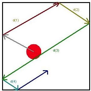 Imagine a ball traveling in a box ; the ball represents a moving molecule. Every time the ball hits a wall, a collision occurs and the direction of the ball changes (Figure 1). The ball hits the wall five times, causing five collisions. Between every two consecutive collisions, the ball travels an individual path. It travels a total of four paths between the five collisions; each path has a specific distance, d. The mean free path, $\lambda$, of this ball is the average length of all four paths.
Source: mean free path ↩︎ -
this is one of two models. ↩︎